Real questions, worked through.
One completed engagement. One early-stage extension, already underway.
Virtual biomarkers
Some of the most useful signal in a tissue section isn't visible to a pathologist's eye, and was never scorable in tools like HALO or Visiopharm: there's no stain, no threshold, no established protocol for it. A model trained on the right data can learn to recognize these features directly from morphology. We call them virtual biomarkers: patterns a trained algorithm can detect and quantify that traditional quantitative-pathology tools can't, because those tools were built to score what's already visible and named, not to find what isn't.
In practice, that means some questions which have historically required building an elaborate multiplex panel just to make a feature visible can instead be answered directly from morphology, with a much simpler wet-lab workflow behind it. Two examples below.
A 5-plex workflow, answered with one morphology model and one serial section
A gene-therapy program needed to identify specific renal tubule populations and confirm therapeutic agent delivery at the level of individual tubules. The standard path to that answer is a 5-plex workflow: a 4-plex multiplex immunofluorescence (mIF) panel to identify and classify the tubule features, plus fluorescence in situ hybridization (FISH) to localize the therapeutic agent's mRNA.
We built a morphology-based algorithmic model that identifies the same renal tubule populations directly from H&E, a virtual biomarker standing in for the 4-plex mIF panel, and paired it with a single serial-section IHC/FISH read to localize the therapeutic agent. One model and one additional section did the work the 5-plex panel was built to do.
No mIF panel to design, optimize, and validate before a single sample could be read, a step that typically adds significant time and cost on its own. The program got tubule-level, quantitative data on both feature identification and therapeutic delivery, faster and at a fraction of the panel-development cost.
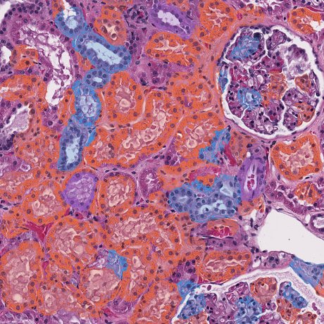
H&E section: tubules annotated and typed directly from morphology.
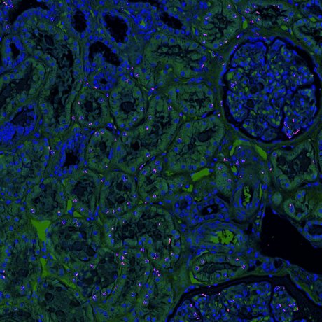
Adjacent serial section, rendered to localize vector-genome signal; puncta simulated for illustration.
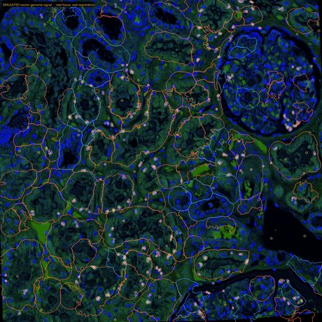
Registered and assigned: detected puncta circled, tubule type carried over from the H&E.
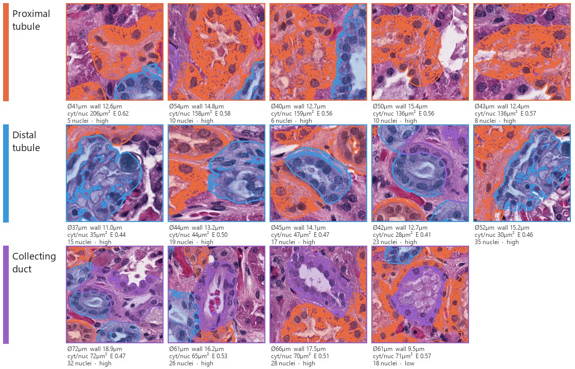
Highest-confidence exemplars per class, with the measurements behind each call. Typing is a transparent, additive score over features that mirror published renal-pathology criteria, not a black box. Cytoplasm-to-nucleus ratio alone separates proximal from distal tubules by roughly 2×, consistently.
Tissue morphology and tubule typing above are real model output on representative tissue. Vector-genome puncta in panels 2–3 are simulated for illustration, not measured signal from a named program.
Where this is headed: liver, without fighting autofluorescence
Liver is a difficult tissue for multiplex immunofluorescence: native autofluorescence from lipofuscin and other endogenous compounds degrades signal-to-noise across stains, which makes mIF panels hard to build and read. Even once a panel works, cell-type identification in liver tissue is laborious.
We're extending the same morphology-based approach to liver: a model that identifies cell populations from morphological features alone, a virtual biomarker for cell type, overlaid with a single serial section of IHC or FISH in place of a full mIF panel. This is an early-stage capability we're actively developing, not a completed engagement, and we're being explicit about that here.
A workflow that never has to fight autofluorescence in the first place, because it isn't built on immunofluorescence, and a laborious multi-stain cell-typing process collapsed into one model and one serial section.