Purpose-built computational pathology.
Not a one-size-fits-all model.
Wet-lab tissue services meet custom computational models, surfacing virtual biomarkers. We train computer vision algorithms to detect "human-non-observable-features" in morphological data, bypassing the need for IHC, IF, and ISH labeling. Any assay that typically requires a pathologist's annotation can be automated. Go beyond traditional subscription-based services and elevate your custom pathology analysis pipeline.
Four capabilities.
Our key differentiator. A two-day proof-of-concept model with a ten-day path to full automated analysis deployment, built, QC'd, and overseen by a computational pathologist.
H&E, special stains, IHC, IF, mIF, ISH/FISH, pathology consultation, and quantitative image analysis.
Custom tools for high-resolution spatial and morphological analysis, generated and interpreted under one roof.
Computational screening of digital biobank slides to enrich rare-subtype sample cohorts before wet-lab confirmation. You may already own the uncharacterized, rare samples.
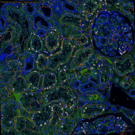
We replaced a 5-plex mIF/FISH workflow with an H&E and serial IHC stain using virtual biomarkers, saving thousands of dollars per sample. We built an H&E based liver cell typing classifier that saved our customer from running costly mIF panels in liver, known for high levels of autofluorescence. See how we've approached real research questions.
See our approach →